PRODUCT DEVELOPMENT
TokeOni™
(also known as:AkaLumine-HCI)Kurogane Kasei is an authorized manufacturer of TokeOni.
Should you require our services for clinical studies, research, or other purposes, please feel free to contact us.
With Kurogane Kasei as an authorized manufacturer…
- Large volume sales are possible.
- We offer reasonable prices.
What is TokeOni?
TokeOni is a luciferin analog that exhibits near-infrared emission (λmax = 675 nm, Figure 1).
TokeOni emits light upon reacting with firefly luminescent enzymes such as Fluc and Akaluc. Thus, it can be used for imaging luciferase-expressing cells, as well as for reporter assays involving luciferase gene transfer.
*TokeOni was developed by Professor Maki of the University of Electro-Communications. Our company has achieved mass production.
Figure 1. Emission spectrum of D-Luciferin and TokeOni
Features of TokeOni
- Near-infrared luminescence is advantageous for observation of deep tissues due to its high biological penetration ability
- Can be used in place of D-luciferin in Fluc systems such as Luc2
- Using the specialized enzyme Akaluc, measurement for deep tissue imaging is more sensitive than D-luciferin×Fluc (Firefly BLI (Bio-Luminescence Imaging))
- What is specialized enzyme Akaluc?
- Akaluc is an artificial enzyme that was developed by a joint research group led by Dr. Atsushi Miyawaki, Team Leader of the Laboratory for Cell Function Dynamics at the RIKEN Center for Brain Science, and Dr. Satoshi Iwano, a Special Postdoctoral Researcher. Its combination with TokeOni substrate is highly compatible and is being used as a tool for deep tissue bioluminescence imaging, i.e., the Aka-BLI system.
RIKEN press release ⇒ https://www.riken.jp/press/2018/20180223_1/
Contact us for inquiries on Akaluc ⇒ https://dna.brc.riken.jp/ja/gene_analysis/inf0039j
Aka-BLI bioluminescence imaging system
-
- 1. Even a small number of cells that could not be detected with Firefly-BLI, can be visualized
- Luc2- and Akaluc-expressing cells were introduced into mice by tail vein injection, at 10 cells/100 cells each.
D-luciferin and TokeOni were then administered by tail vein injection. (Figure 2)
Both groups of 10 cells and 100 cells were barely visible with Firefly-BLI, but clearly detectable with Aka-BLI.
Figure 2. Visualization of luciferase-expressing cells: 10 cells (left)/100 cells (right)
(Images provided by Dr. Takahiro Kuchimaru, Associate Professor, Jichi Medical University) -
- 2. Actualizing ultra-sensitive brain imaging
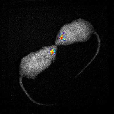
- While imaging of the brain with Firefly-BLI is extremely difficult due to the influence of the blood-brain barrier (BBB), Aka-BLI has a detection sensitivity of approximately 1,400 times more than the former, making it highly sensitive for brain imaging. In addition, observing moving images in real time is possible (Figure 3).
Figure 3. Brain imaging of two freely moving mice
(Image provided by Dr. Satoshi Iwano, RIKEN) -
- 3. Real-time observation of marmoset brain is possible
- Brain visualization with Aka-BLI has been achieved in not only mice and other small animals, but also in small primates such as marmosets (Figure 4).
Here, both the actual moving marmoset and the emission from its brain can be detected at the same time.
Figure 4. Brain imaging of freely moving marmoset
(Image provided by Dr. Satoshi Iwano, RIKEN)
TokeOni’s examples of use
TokeOni should be prepared in water or physiological saline when administering to animals.
It can emit light at sites where luminescent enzymes (Luc2 and Akaluc) are present.
*Cautionary points
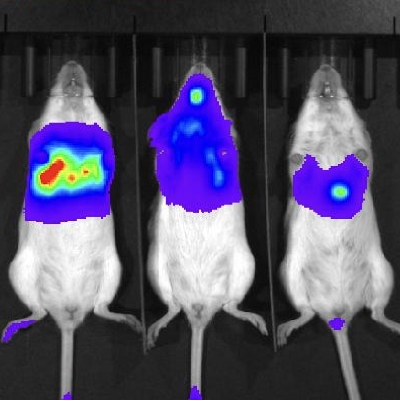
When TokeOni is introduced into a living body, it is often confirmed that the liver autofluoresces. There are individual differences depending on the animal, but please be careful when measuring the emission brightness near the liver.
Detection of lung metastatic cancer (site: lung, enzyme: Luc2)
Kuchimaru, T., et al, “A luciferin analogue generating near-infrared bioluminescence achieves highly sensitive deep-tissue imaging”. Nat. Commun. 7, 11856(2016)
Visualization of brain-derived neurotrophic factor (BDNF) (site: brain, enzymes: Luc2)
M. Fukuchi, et. al., “Visualization of activity-regulated BDNF expression in the living mouse brain using non-invasive near-infrared
bioluminescence imaging”. Mol. Brain. 13, 112(2020)
M. Fukuchi, et. al., “Visualizing changes in brain-derived neurotrophic factor (BDNF) expression using bioluminescence imaging in
living mice”. Sci. Rep., 7, 4949(2017)
Detection of adeno-associated virus (site: brain (mouse), brain (marmoset), enzyme: Akaluc)
/ Detection of c-fos gene expression (site: brain, enzyme: Akaluc)/ Detection of a single tumor cell (site: lung, enzyme: Akaluc)
Visualization of pluripotent stem cells (site: subcutaneous, enzyme: Akaluc)
Martin, R.M., et. al., “Improving the safety of human pluripotent stem cell therapies using genome‐edited orthogonal safeguards, Nat. Commun. 11,2713 (2020)
Visualization of myelodysplastic syndrome (MDS) cell line (site: whole body, enzyme: Akaluc)
Zhong, C., et. al., “Efficacy of the novel tubulin polymerization inhibitor PTC‐028 for myelodysplastic syndrome”,
Cancer Sci. 12, 4336–4347 (2020)
Safety of TokeOni
Information on safe handling and storage of TokeOni, and its chemical composition will be provided through the “SDS (Safety Data Sheet).”
Request for quotation/Inquiry form
If you have any questions about TokeOni, or would like to request for a quotation, please contact us using the inquiry form below.